

New Fluorophore Technology Promises Faster, Clearer Cellular Imaging
Researchers Develop Photocleavable “CLEAR” Probe for Advanced Live Cell Microscopy
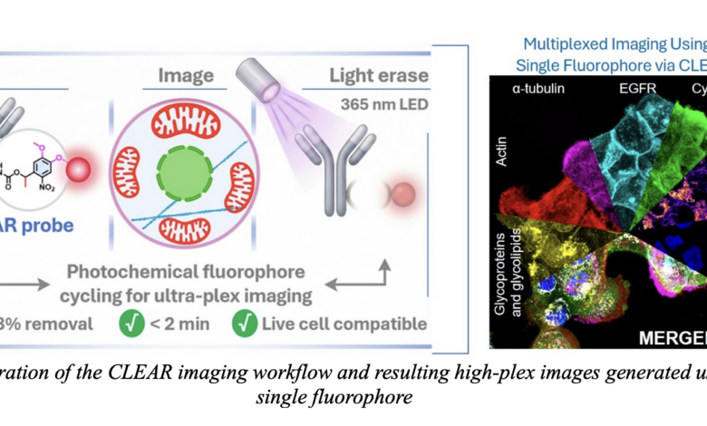
Scientists have developed a breakthrough imaging technology that could significantly improve the way living cells are studied under microscopes. The innovation, known as the “CLEAR probe,” enables researchers to capture high-quality cellular images while removing excess fluorescent signals that often interfere with imaging accuracy.
The new system works through a photocleavable fluorophore mechanism, where unwanted fluorescent markers can be removed using 365 nm LED light exposure in less than two minutes. According to the research visuals, the technique achieves over 98 percent signal removal while remaining compatible with live cells, making it highly useful for biological and medical research.
The CLEAR probe is designed to simplify multiplexed imaging — a process that allows scientists to observe multiple biological structures simultaneously. Traditional fluorescence imaging often suffers from signal overlap and background interference, limiting the number of cellular components that can be analyzed at once. The new approach aims to overcome these limitations by enabling repeated cycles of labeling, imaging, and light-triggered signal removal.
Images released by researchers demonstrate how different cellular structures such as actin, EGFR, mitochondria, tubulin, and cytokeratin can be visualized together in vivid colors. The merged cellular image highlights the technology’s potential for high-resolution mapping of complex biological systems.
Researchers believe the advancement could play a crucial role in cancer biology, neuroscience, drug discovery, and precision medicine, where accurate imaging of cellular interactions is essential. Because the process is compatible with living cells, scientists can also monitor dynamic biological activity in real time without causing major cellular damage.
The use of low-intensity LED light instead of harsher chemical treatments is another major advantage, potentially reducing toxicity and preserving delicate biological samples. The technology may also help laboratories reduce imaging time and improve efficiency in large-scale biomedical studies.
Experts say innovations in multiplex fluorescence imaging are becoming increasingly important as researchers seek deeper insights into disease mechanisms at the molecular level. The CLEAR probe system could eventually support next-generation diagnostic platforms and advanced biomedical imaging tools used in hospitals and research institutes worldwide.
The development reflects growing efforts in the scientific community to combine precision imaging with faster and safer laboratory techniques, opening new possibilities for understanding human cells and complex diseases with unprecedented detail.